Protein compatible hydrogel nanofilms

Hydrogel network synthesis
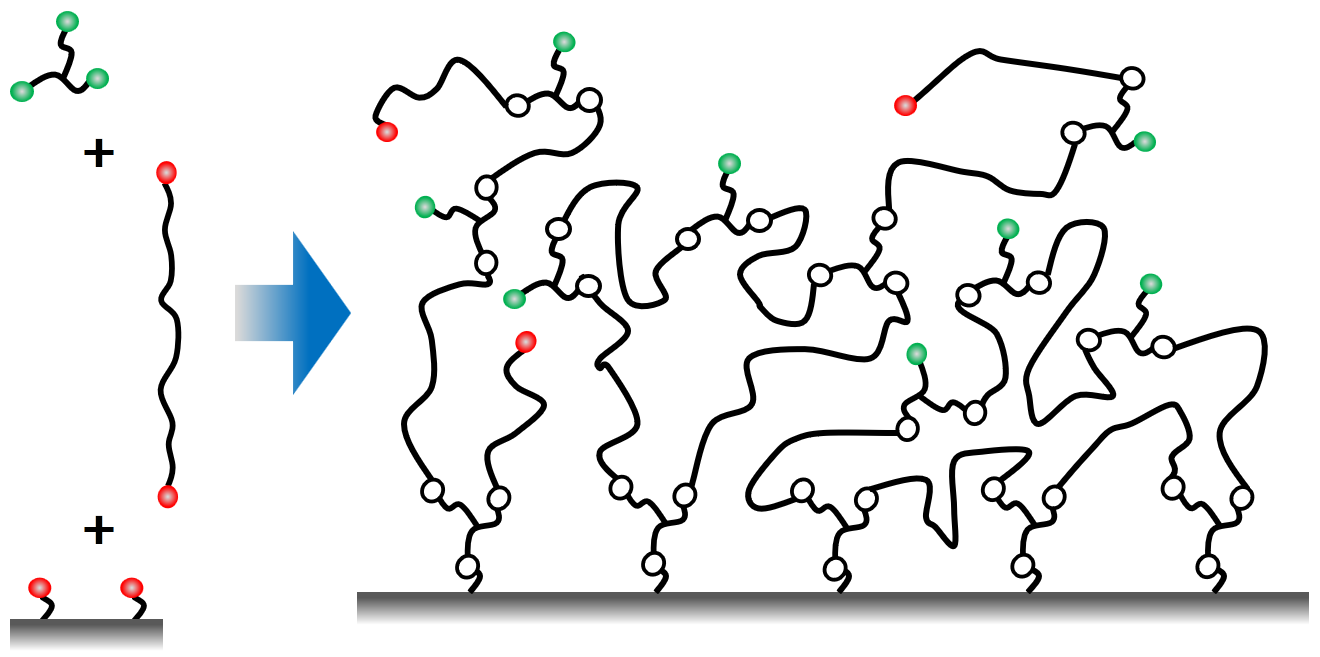
Hydrogel network formation
© Fraunhofer IAP
Formation of a network from long bifunctional branch molecules and trifunctional linkers
Extended scheme for network formation
© Fraunhofer IAP
Formation of a network from long bifunctional branch molecules, trifunctional linkers, and hetero-bifunctional modifiers

Proteins often denature if they interact with surfaces. The so-called unspecific interactions between proteins and surfaces are usually responsible for the denaturation. Looking at a polymer surface and a protein on a molecular scale shows that the many possible interaction sites gives a good reasoning for this effect.  

These unwanted interaction are extremely small with hydrogels.A hydrogel network with a sufficient pore size can host proteins. These hydrogels provide proteins with their natural environment which keeps them in their functional state.

The networks can be constructed from long bifunctional branch molecule which are connected to each other and to the surface by trifunction linkers as shown in the scheme. Depending on the details of how the reaction is carried out some of the functional groups remain at the end and can be used for the immobilization. In this way the concentration of the immobilization sites and also network desity can be tailored (Hydrogel Nanofilms for Biomedical Applications)

Adding additional components to the reaction can modify the simple scheme and provides even more freedom for the design the structure of the network. For example, a hetero-bifunctional modifier get incorporated into the network if one of the two functional groups can take part in the network formation reaction. With this scheme the concentration of functional groups for immobilzation can be tuned or a new type of group can be introduced. The example in the scheme shows a network which is formed from diamino-PEG and a triacid chloride. An amino acid (alanine) also reacts with the acid chloride and increases the concentration of carboxylic acid groups in the network.   
 

Properties of the hydrogel networks

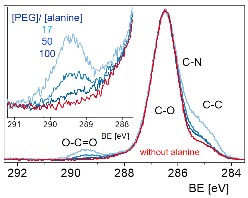
XPS alanine-PEG network
© Fraunhofer IAP
XP C1s spectra of an alanine modified PEG network with varying alanine concentration.
PEG network with alanine, carboxylic acid groups labelled
© Fraunhofer IAP
Fluorescence spectra of carboxylic acid goups labelled with dansysl cadaverine contained in an alanine modified PEG network with varying alanine concentration..
Large proteins interacting inside the network
© Fraunhofer IAP
A network immobilizes sequencielly straptavidin, biotinylated protein A, and an FITC-labelled antibody. Fluorescence of the FITC after the reaction with biotin-containing and a biotine-free network.

The technique for the preparation of hydrogel films is very versatile. Many parameters and properties of the hydrogel can be adjusted to the needs of a specific application:

film thickness

network density

immobilization chemistry

density of the immobilization sites

There is an almost unlimited range of substrate materials to be coated with the hydrogel. For the immobilization of proteins, a variety of techniques can be utilized as for example the classic biotin-streptavidin way, the click chemistry, or the EDC/NHS technique.  The concentration of immobilization sites can be adjusted to the requirements in a wide range. Hydrogels with a varying carboxylic acid group concentration, for example, are prepared by adding different amounts of alanine to the reaction solution. The concentration of COOH groups can be determined by XPS and by fluorescence labelling.

A hydrogel that can be used for biotin-streptavidin based conjugation has biotin coupled to the network e.g. via a biotinylated amino-PEG. After immobilizing streptavidine a biotinylated protein a can be added which, finally, is tagged using an antibody with a fluorescent label (FITC). The fluorescence is much larger than expected from a flat substrate and suggests a stacked three-dimensional immobilization. If the same immobilization sequence is applied to a biotin-free network the final fluorescence signal is very small.

Applications

These hydrogels provide proteins with their natural environment which keeps them in their functional state. Thus, such gels can be used to study the function of the immobilized proteins or to exploit the function in a diagnostic device. The layers prepared from the hydrogels are thin to allow a rapid diffusion into the gel and out of it.

The technology developed at the Fraunhofer IAP allows the efficient production of surface bound hydrogel nanofilms. Requests of a specific application can be incorporated to detail the preparation procedure. 3D-hydrogels coupled to a polymer surface can provide new opportunities for medical diagnostics and biological research. Beside the application in biological sensors and diagnosis devices there are many more fields of application. One could think of investigating the interaction with biological cells or of preparing protein resistant surfaces.